
Advanced Mycological Applications of Melzer Reagent: Chemistry, Diagnostics, and Taxonomic Interpretation
Melzer Reagent—commonly called Melzer’s Solution—is one of the foundational iodine-based reagents in fungal microscopy, especially for the identification of amyloid and dextrinoid reactions in spores, asci, hyphae, and tissue structures. Originally formulated by Vilem Melzer, this reagent continues to be indispensable in classical and modern taxonomy, appearing in research manuals from Cornell University (https://plantpathology.cals.cornell.edu), Oregon State University Mycology Lab (https://bpp.oregonstate.edu), and USDA Forest Service fungal guides (https://www.fs.fed.us).
Today, Melzer Reagent remains central not only for observational taxonomy but also for linking morphological traits to molecular phylogenetics, as supported by studies indexed in NCBI (https://www.ncbi.nlm.nih.gov) and sequencing frameworks integrated into the National Human Genome Research Institute (https://www.genome.gov).
Historical Evolution of Melzer Reagent
The history of Melzer Reagent is tightly integrated with European mycology and early North American fungal taxonomy, highlighted in archives from Harvard University Herbaria (https://huh.harvard.edu) and the Smithsonian National Museum of Natural History (https://naturalhistory.si.edu).
Pre-Melzer Iodine Techniques
Before Melzer’s formulation, mycologists relied on Lugol’s iodine or potassium iodide solutions described in 19th-century protocols conserved in Library of Congress science archives (https://www.loc.gov). These older solutions lacked:
-
Sufficient tissue clearing
-
Reliable coloration stability
-
Strong contrast enhancement
This limitation drove interest in synthesizing a superior clearing and staining reagent.
Development of the Chloral Hydrate Formula
The addition of chloral hydrate fundamentally changed fungal microscopy. As documented in chemical safety guidelines from NIH PubChem (https://pubchem.ncbi.nlm.nih.gov), chloral hydrate acts as:
-
A clearing agent
-
A refractive index stabilizer
-
A solvent for iodine penetration
This allowed reproducible amyloid and dextrinoid reactions across thousands of Basidiomycota species documented in USDA fungal host databases (https://nt.ars-grin.gov/fungaldatabases).
Chemical Composition and Mode of Action
Melzer Reagent generally contains:
-
Iodine (I₂)
-
Potassium iodide (KI)
-
Chloral hydrate
-
Distilled water
Chemical behavior of iodine complexes is explored deeply in materials from University of Illinois Chemistry Department (https://chemistry.illinois.edu) and University of California Davis Biology Program (https://biology.ucdavis.edu).
Amyloid Reaction Mechanism
Amyloid reactions involve iodine binding to β-linked polysaccharides in the fungal cell wall. Research into the structural diversity of fungal glucans appears frequently in papers archived in NCBI PMC (https://www.ncbi.nlm.nih.gov/pmc).
The blue-black amyloid color arises from a reaction analogous to the iodine-starch complex described in classical chemistry courses at MIT (https://chemistry.mit.edu).
Dextrinoid Reaction Mechanism
Dextrinoid reactions (red-brown) indicate:
-
Modified β-glucan branching
-
High densities of mannoproteins
-
Unique spore ornamentation chemistry
These concepts appear in plant pathology training modules from Penn State Extension (https://extension.psu.edu) and in fungal ultrastructure resources at the University of Minnesota CBS (https://cbs.umn.edu).

Diagnostic Value in Mycology
The primary reason for the reagent’s widespread use is its taxonomic precision, supported in US government and academic fungal identification manuals such as:
-
USFS Field Guides (https://www.fs.usda.gov)
-
USDA NRCS PLANTS (https://plants.usda.gov)
-
University of Georgia Plant Pathology (https://plantpath.caes.uga.edu)
Spore Ornamentation and Wall Morphology
Melzer Reagent reveals:
-
Amyloid ornamentation in Russulales
-
Dextrinoid warts in Boletales
-
Inamyloid smooth spores in many Agaricales
Fungal microscopic atlases from University of Michigan Herbarium (https://lsa.umich.edu/herbarium) provide extensive examples of how these reactions support species-level identification.
Ascus Apex Iodine Reaction
In Ascomycota, Melzer Reagent is used to distinguish:
-
Iodine-positive asci (unitunicate-operculate)
-
Iodine-negative asci
-
Variably amyloid apical rings
Studies of ascus iodine reactions are referenced in Berkeley Mycology Teaching Laboratory (https://nature.berkeley.edu).
Comparison With Lugol’s and Other Iodine Reagents Lugol’s Solution
Lugol’s is widely used in plant pathology training from University of Florida IFAS (https://edis.ifas.ufl.edu), but lacks:
-
Chloral hydrate (critical for clearing)
-
High-resolution contrast
-
Reliable amyloid reaction stability
IKI (Iodine Potassium Iodide)
IKI is used in lichenology, particularly in research from University of Vermont Field Naturalist Program (https://www.uvm.edu), but Melzer Reagent still provides superior differentiation of dextrinoid structures.
Modern Applications: From Classical Morphology to Genomic Integration
Though originally a classical morphological tool, Melzer Reagent is now integrated with:
-
DNA barcoding datasets from BOLD (https://www.boldsystems.org)
-
ITS sequencing frameworks described by the US National Institutes of Health (https://www.nih.gov)
-
Phylogenomic pipelines explored in computational training by Carnegie Mellon University (https://www.cmu.edu)
This “morpho-molecular duality” is increasingly emphasized by experts publishing through NCBI Bookshelf (https://www.ncbi.nlm.nih.gov/books).
5.1 Voucher Specimen Documentation
Modern fungal vouchering guidelines from the USGS Biological Resources Division (https://www.usgs.gov) recommend recording:
-
Amyloid reactions
-
Dextrinoid reactions
-
Inamyloid tissues
-
Microscopic ornamentation micrographs
Experimental Protocol: Best Practices
Guidelines harmonize with laboratory safety protocols published by:
-
CDC NIOSH (https://www.cdc.gov/niosh)
-
OSHA Laboratory Standard (https://www.osha.gov/laboratory-safety)
-
EPA hazardous waste management (https://www.epa.gov/hw)
Slide Preparation
-
Place a thin fragment of fungal tissue on a slide.
-
Add 1–2 drops of Melzer Reagent.
-
Allow clearing for 20–60 seconds.
-
Add coverslip gently, avoiding air bubbles.
-
Observe under 400–1000× magnification.
Optical adjustments are reviewed in microscopy teaching resources from University of Washington Biology (https://www.biology.washington.edu).
Reaction Timing
The amyloid and dextrinoid reactions stabilize within 10–20 seconds, but the reagent continues clearing tissue for several minutes.
Image Documentation
High-quality imaging protocols are detailed in NIH ImageJ user guides (https://imagej.nih.gov/ij).

Chemical Stability, Storage & Shelf-Life
According to chemical guidelines from DOE Environment, Health, Safety & Security (https://www.energy.gov/ehss), Melzer Reagent should be:
-
Stored in amber glass
-
Kept away from light
-
Maintained at stable room temperature
-
Disposed according to iodine chemical waste procedures
Advanced Interpretation of Amyloid and Dextrinoid Patterns
Basidiomycota
-
Amanita spp.: Clear amyloid reaction in spores
-
Lactarius spp.: Dextrinoid ornamentation
-
Russula spp.: Strongly amyloid spore amyloidity
These patterns are part of taxonomic keys in USFS North American Fungi Guides (https://www.fs.fed.us/wildflowers).
Ascomycota
-
Amyloid apical rings in Helotiales
-
Negative reactions in Xylariales
-
Variable iodine reaction in lichenized fungi (as described by University of Wisconsin Botany: https://botany.wisc.edu)
Zygomycota and Mucoromycotina
Mostly inamyloid, but Melzer Reagent assists in tissue clearing for morphological measurements documented in University of Kentucky Biology (https://bio.as.uky.edu).
Reagent Limitations and Considerations
Despite its utility, Melzer Reagent has limitations:
-
Chloral hydrate regulations
-
Batch-to-batch variation
-
Potential reagent crystallization
-
Occasional false negatives
Analyses of reagent performance variability are discussed in USDA Northern Research Station studies (https://www.nrs.fs.usda.gov).
Conclusion
Melzer Reagent remains one of the most technically valuable, taxonomically essential, and scientifically validated reagents in mycology. Its ability to detect amyloid and dextrinoid reactions connects classical microscopy with modern phylogenomic research, supported by decades of publications, field manuals, academic courses, and federal research documentation from numerous .edu and .gov institutions.
It continues to serve as a critical tool for:
-
Fungal taxonomy
-
Fungal identification in environmental surveys
-
Microscopic characterization of spores and asci
-
Integrating morphology with molecular data
-
High-resolution microscopy workflows

StressMarq
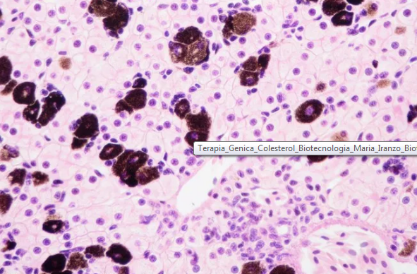
Gene therapy to lower cholesterol
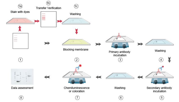
Western Blot Protocols (part 3) – Antibody Incubation & Gel Visualization
About Author
